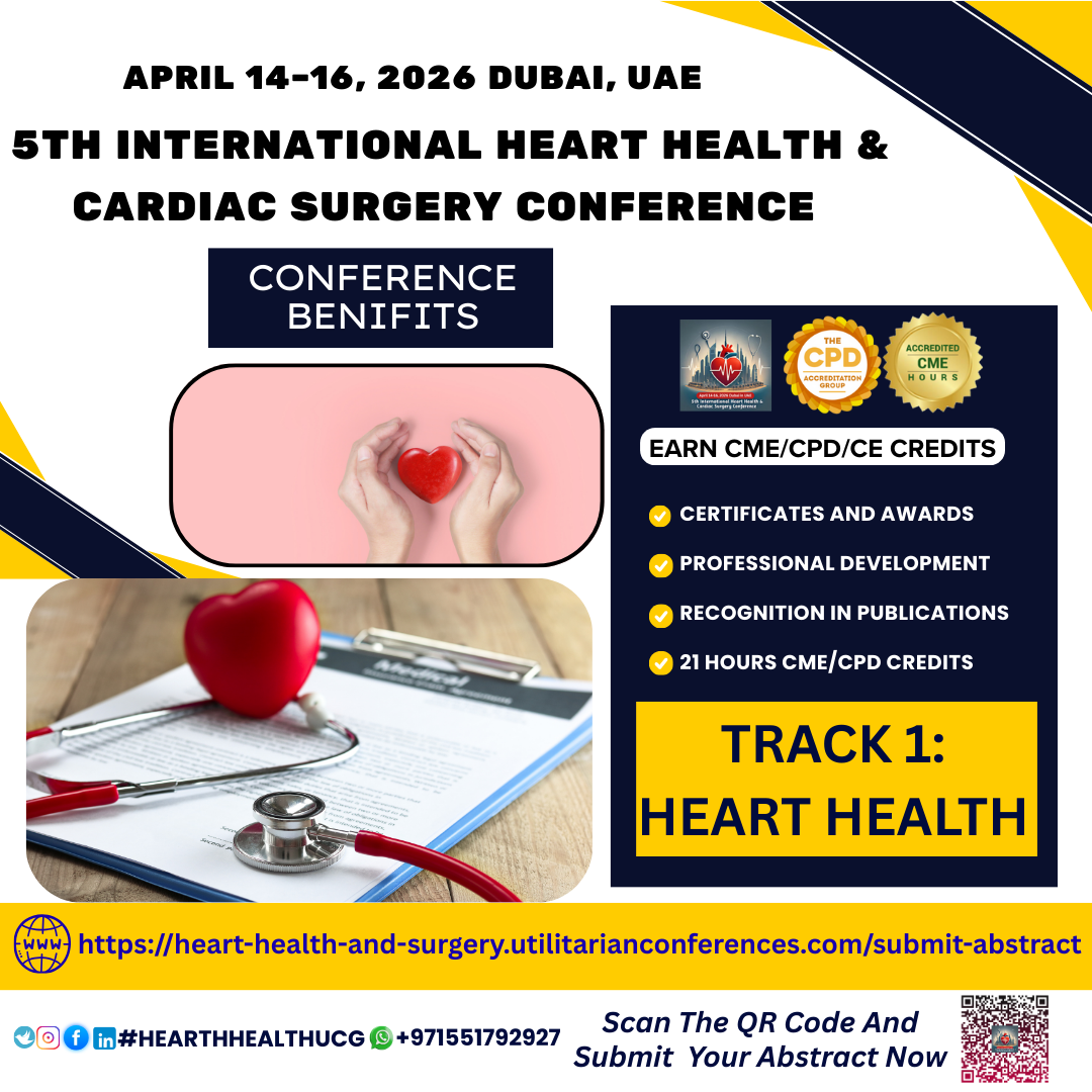
Heart Health is a broad and essential topic that refers to maintaining the...
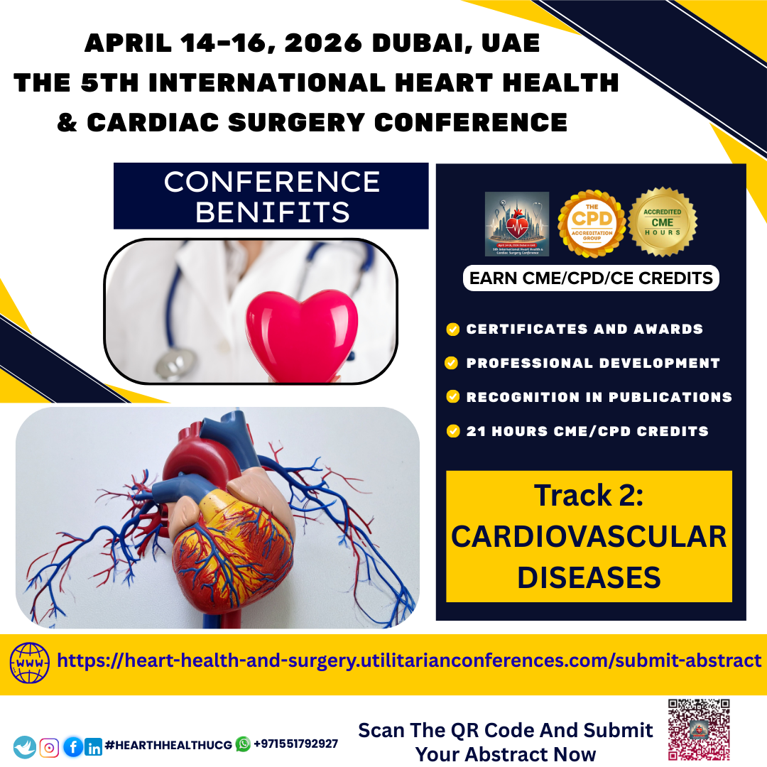
Cardiovascular diseases (CVDs) are the leading cause of death globally,
claiming an estimated 17.9...
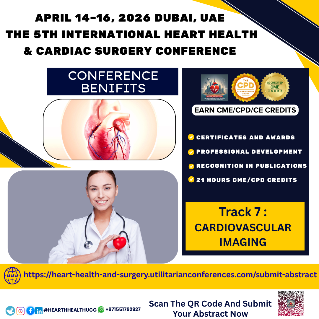
Heart disease remains one of the leading causes of death worldwide. Yet, many heart conditions can be detected early—and managed effectively—thanks to a powerful set of tools known as cardiovascular
imaging. These non-invasive and minimally invasive techniques allow physicians to "see" the heart and blood vessels in action, helping diagnose diseases, monitor progress, and guide treatment decisions.
Cardiovascular imaging refers to a group of medical imaging techniques used to visualize the structure and function of the heart and vascular system. It plays a crucial role in detecting abnormalities such as blocked arteries, heart muscle damage, valve disorders, congenital defects, and more.
Whether you're experiencing chest pain, shortness of breath, or simply undergoing a routine check-up, your doctor may recommend one or more imaging tests depending on your symptoms and risk factors.
Common Types of Cardiovascular ImagingOften the first-line imaging test, an echocardiogram uses ultrasound waves to create real-time images of the heart. It can show how well your heart is pumping, whether your valves are working properly, and if there’s fluid around the heart.
Stress Echo: Assesses heart function under stress.
Transesophageal
Echo (TEE): Offers more detailed views via an ultrasound probe passed
into the esophagus.
Fast, non-invasive.
Helps evaluate risk of heart attack.
3. Cardiac MRI (Magnetic Resonance Imaging)
Cardiac MRI provides high-resolution images of the heart’s soft tissues and is valuable for evaluating cardiomyopathies (diseases of the heart muscle), congenital heart disease, and damage after a heart attack.
No radiation.
Highly detailed anatomy and function analysis.
4. Nuclear Cardiology (SPECT & PET)These tests use small amounts of radioactive material to assess blood flow to the heart muscle—often used for diagnosing coronary artery disease.
·
SPECT: Widely used for stress testing.
·
PET: Offers higher resolution and greater accuracy in certain cases.
Used during cardiac catheterization procedures, these imaging methods give detailed pictures of the inside of blood vessels, often helping guide stent placement.
The right imaging test can be the difference
between early detection and a missed diagnosis. Imaging helps:
·
Identify heart disease before symptoms arise
·
Guide treatment strategies—whether medication, surgery, or intervention
·
Monitor the effectiveness of therapie
Reduce the risk of sudden cardiac events
Thanks to advances in artificial intelligence, 3D imaging, and machine learning, cardiovascular imaging is becoming faster, more accurate, and more predictive than ever before. These tools are not only diagnosing disease but also helping predict future cardiovascular events—a leap forward in preventive cardiology.